fbpx

Our Services

Schedule Appointment
780-444-6407

Learn More About Our Services

Eyewear Makeovers

Comprehensive Eye Exams

Pediatric Eye Exams

Vision Therapy


Eye Emergencies (Pink/Red Eyes)

Management of Ocular Diseases


Treatment Of Vision-Related Learning Issues In Children 2

Treatment Of Vision-Related Learning Issues in Children

Edmonton Optometrist

Glaucoma Testing


Pediatric Eye Exams

As a parent, you may wonder whether your preschooler has a vision problem or when a first eye exam should be scheduled.

Eye exams for children are extremely important. Experts say 5 to 10% of preschoolers and 25% of school-aged children have vision problems. Early identification of a child’s vision problem is crucial because, if left untreated, some childhood vision problems can cause permanent vision loss.


When should kids have their eyes examined?

According to the Alberta Optometry Association (AOA), infants should have their first comprehensive eye exam at 6 months of age. We recommend that children should receive additional eye exams yearly.

Children who need eyeglasses or contact lenses should be examined annually or according to their eye doctor’s recommendations.

Early eye exams also are important because children need the following basic visual skills for learning:

  • Near vision
  • Distance vision
  • Eye teaming (binocularity) skills
  • Eye movement skills
  • Focusing skills
  • Peripheral awareness
  • Eye/hand coordination

Because of the importance of good vision for learning, some states require an eye exam for all children entering school for the first time.

Scheduling your child’s eye exam.

Your family doctor or pediatrician likely will be the first medical professional to examine your child’s eyes, however, even if your pediatrician has checked your child’s eyes we still recommend routine exams starting at 6 months of age. Eye doctors have specific equipment and training to help them detect and diagnose potential vision problems and are able to ensure your child’s vision development is on track. .

When scheduling an eye exam, choose a time when your child is usually alert and happy. Specifics of how eye exams are conducted depend on your child’s age, but an exam generally will involve a case history, vision testing, determination of whether eyeglasses are needed, testing of eye alignment, eye tracking and focusing skills, an eye health examination and a consultation with you regarding the findings.

After you’ve made the appointment, you may be sent a case history form by email, or you may be given one when you check in at the doctor’s office. The case history form will ask about your child’s birth history (also called perinatal history), such as birth weight and whether or not the child was full-term. Your eye doctor also may ask whether complications occurred during the pregnancy or delivery. The form will also inquire about your child’s medical history, including current medications and past or present allergies.

Be sure to tell your eye doctor if your child has a history of prematurity, has delayed motor development, engages in frequent eye rubbing, blinks excessively, fails to maintain eye contact, cannot seem to maintain a gaze (fixation) while looking at objects, has poor eye tracking skills or has failed a pediatrician or pre-school vision screening.

Your eye doctor will also want to know about previous ocular diagnosis and treatments involving your child, such as possible surgeries and glasses or contact lens wear. Be sure you inform your eye doctor if there is a family history of eye problems requiring vision correction, such as nearsightedness or farsightedness, misaligned eyes (strabismus) or amblyopia (“lazy eye”).

Eye testing for infants.

It takes some time for a baby’s vision skills to develop. To assess whether your infant’s eyes are developing normally, your eye doctor may use one or more of the following tests:

Tests of pupil responses evaluate whether the eye’s pupil opens and closes properly in the presence or absence of light.
“Fixate and follow” testing determines whether your baby can fixate on an object (such as a light) and follow it as it moves. Infants should be able to perform this task quite well by the time they are 3 months old.
Preferential looking involves using cards that are blank on one side with stripes on the other side to attract the gaze of an infant to the stripes. In this way, vision capabilities can be assessed.

Eye testing for preschool children.

Pre-school children can have their eyes thoroughly tested even if they don’t yet know the alphabet or are too young or too shy to answer the doctor’s questions. Some common eye tests used specifically for young children include:

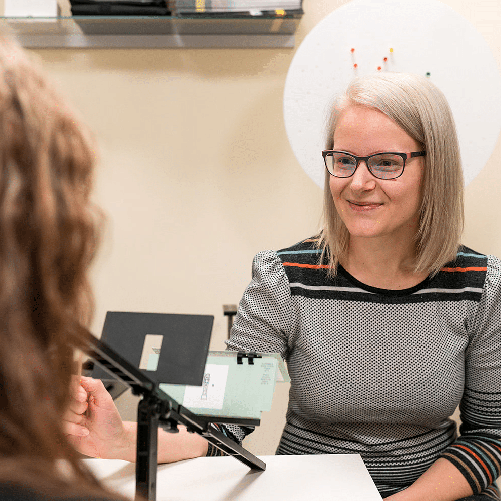
LEA Symbols for young children are similar to regular eye tests using charts with letters, except that special symbols in these tests include an apple, house, square and circle.

Retinoscopy is a test that involves shining a light into the eye to observing how it reflects from the retina (the light-sensitive inner lining of the back of the eye). This test helps eye doctors determine the child’s eyeglass prescription.

Random Dot Stereopsis uses dot patterns to determine how well the two eyes work as a team.

Eye and vision problems that affect children.

Besides looking for nearsightedness, farsightedness and astigmatism (refractive errors), your eye doctor will be examining your child’s eyes for signs of these eye and vision problems commonly found in young children:

Amblyopia: Also commonly called “lazy eye,” this is decreased vision in one or both eyes despite the absence of any eye health problem or damage. Common causes of amblyopia include strabismus (see below) and a significant difference in the refractive errors of the two eyes. Treatment of amblyopia may include patching the dominant eye to strengthen the weaker eye.
Strabismus: This is misalignment of the eyes, often caused by a congenital defect in the positioning or strength of muscles that are attached to the eye and which control eye positioning and movement. Left untreated, strabismus can cause amblyopia in the misaligned eye. Depending on its cause and severity, surgery may be required to treat strabismus.
Convergence insufficiency: This is the inability to keep the eye comfortably aligned for reading and other near tasks. Convergence insufficiency can often be successfully treated with vision therapy, a specific program of eye exercises.
Focusing problems: Children with focusing problems (also called accommodation problems) may have trouble changing focus from distance to near and back again (accommodative infacility) or have problems maintaining adequate focus for reading (accommodative insufficiency). These problems often can be successfully treated with vision therapy or glasses.
Eye teaming problems: Many eye teaming (binocularity) problems are more subtle than strabismus. Deficiencies in eye teaming skills can cause problems with depth perception and coordination.

Vision and learning.
Experts say that 80% of what your child learns in school is presented visually. Undetected vision problems can put them at a significant disadvantage. Be sure to schedule a complete eye exam for your child prior to the start of school.


Source: Eye Exams for Children, article by AllAboutVision.com. ©2009 Access Media Group LLC. All rights reserved. Reproduction other than for one-time personal use is strictly prohibited.

Comprehensive Eye Exams Edmonton

Vision by Design offering comprehensive Eye Exams in Edmonton
Regardless of your age or physical health, it’s important to have regular comprehensive eye exams.

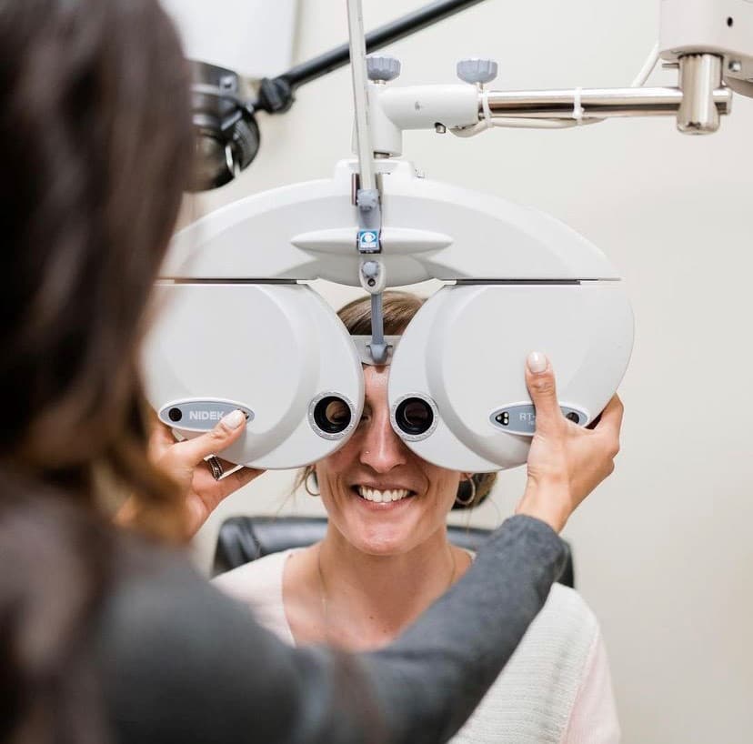
During a complete eye exam, your eye doctor will not only determine your prescription for eyeglasses or contact lenses, but will also check your eyes for diseases of the eyes, assess how your eyes work together as a team and evaluate your eyes as an indicator of your overall health.

A comprehensive eye exam includes a number of tests and procedures to examine and evaluate the health of your eyes and the quality of your vision. These tests range from simple ones, like having you read an eye chart, to complex tests, such as using a high-powered lens to examine the health of the tissues inside of your eyes. Vision by Design Optometry also has specialized equipment that allows us to monitor for diseases such as glaucoma, macular degeneration and cataracts.

We recommend annual eye exams even if your vision is stable, as it allows us to monitor for diseases of the back of the eye, which often do not have any symptoms in the early stages.

Some experts estimate that approximately 5% to 10% of preschoolers and 25% of school-aged children have vision problems. According to the Alberta Optometry Association, all children should have their eyes examined at 6 months of age, and then annually. Annual exams are recommended to monitor your child’s vision and ocular health. Often children will not complain of vision problems and early detection is the key to successful treatment.

Children without vision problems or risk factors for eye or vision problems should then continue to have their eyes examined at least every year throughout school.

Children with existing vision problems or risk factors should have their eyes examined more frequently.

Common risk factors for vision problems include:

  • Premature birth
  • Developmental delays
  • Turned or crossed eyes
  • Family history of eye disease
  • History of eye injury
  • Other physical illness or disease

The Alberta Optometry Association recommends that children who wear eyeglasses or contact lenses should have their eyes examined at least every 12 months or according to their eye doctor’s instructions.

Adults. The Alberta Optometry Association recommends annual exams for all adults if you wear glasses or contact lenses. If you do not wear glasses or contact lenses annual exams are still recommended as they allow us to monitor for diseases of the back of the eye which may not always have symptoms in the early stages. Doctors often recommend more frequent examinations for adults with diabetes, high blood pressure and other disorders, because many diseases can have an impact on vision and eye health.

If you are over 40, it’s a good idea to have your eyes examined every one to two years to check for common age-related eye problems such as presbyopia, cataracts and macular degeneration.

Because the risk of eye disease continues to increase with advancing age, everyone over the age of 60 should be examined annually.

Eye Emergencies

We are always willing to help, should you ever experience an eye emergency. Our office provides emergency services for eye infections, eye injuries and other eye urgencies. State of the art equipment allows us to examine the front surface of the eye and also digitally scan inside the eye for infection or damage. We accommodate many eye emergencies such as:

  • Eye infections
  • Foreign materials stuck in the eyes
  • Eye trauma
  • Scratched eyes
  • Sudden loss of vision in one or both eyes
  • Lost or broken contact lenses or eyeglasses
  • Flashes of light in the vision
  • “Floaters” in the vision
  • Red or painful eyes
  • Dislodged contact lenses
  • Uncomfortable, itchy, or irritated eyes

Studies have shown that an overwhelming number of emergency room visits could have been treated by an optometrist. These ranged from foreign bodies to severe eye allergies to eye infections as the most common reasons for emergency room visits. It is not always necessary to go to an emergency room for eye emergencies. Optometrists are equipped to treat the majority of eye emergencies.

We understand the importance of eye care when you encounter symptoms such as those listed above. These are signs that an immediate evaluation or consultation is necessary – please call us to set one up if you are experiencing an eye emergency of any kind.

Foreign Body Removal

A foreign body is something such as an eyelash, sawdust, sand, or dirt can that gets into the eyes. The main symptom is irritation or pain. Depending on what it is and how the injury happened, the foreign body may pierce the eye and cause serious injury or it may simply go away with no long-term problem.

The foreign object may set off an inflammatory cascade, resulting in dilation of the surrounding vessels and subsequent edema of the lids, conjunctiva, and cornea. If not removed, a foreign body can cause infection.

If anything is stuck in your eye for more than a period of a couple of hours, you must immediately cease all attempts to remove it yourself. Keep in mind that the eyes are an extremely delicate organ and any attempts to try anything extraordinary with them can only have negative and adverse results. If the foreign body you are talking about is not bothering you too much, then you are advised to visit an eye doctor to take care of it. If not you may need to call the emergency service of your region.

If there is a foreign body in your eye, such as a piece of grit, your eye doctor may try and remove it. They will put anaesthetic eye drops in your eye first, in order to numb it and prevent any pain.

If the foreign body is easy to get to, it may be possible to remove it by simply rinsing your eye with water, or by wiping it away with a cotton wool bud or special instrument. However, if this is unsuccessful, your eye doctor may try and remove the foreign body by lifting it out with the tip of a small metal instrument or using a powered bud.

The foreign body could be stuck underneath your upper eyelid, especially if you can feel something there, or you have scratches or grazes (abrasions) on the top half of the transparent outer layer of your eye (cornea). If this is the case, it may be necessary to gently turn your eyelid inside out in order to remove the foreign body.

Once the anaesthetic eye drops have worn off, your eye may feel a bit uncomfortable until your abrasion heals.

Whatever is happening with your eyes or if you suffer or even suspect that a foreign body has penetrated the outer eye layer better go without delay to the nearest treatment center. Doing nothing can lead to loss of vision, premature cataracts and damage to the retina so do not take any chances, delay is dangerous.


Source: Removing a Foreign Body from Your Eye, article by CareEyeEasy.com. All rights reserved. Reproduction other than for one-time personal use is strictly prohibited.

Treatment of Vision-Related Learning Problems in Children

The connection between good vision and success in school is undisputed. Experts say that about 80 percent of what a student learns in school is information that is presented visually. We live in a visual world. So good vision is essential for a student of any age to reach his full potential and find success in the school setting.

If your child is not succeeding in school, ruling out vision problems should be one of your first steps. Our doctors have the skills and expertise to identify if a vision problem is interfering with your child’s ability to access information and participate fully in school and in after-school activities.
Your child may be nearsighted (can’t see far away objects like a blackboard), farsighted (can’t see objects that are close, such as reading a book) and have astigmatism (a blurring caused by the eyes inability to focus light appropriately).

Watch for these symptoms in conjunction with school challenges:

  • Headaches or eye strain
  • Blurred vision or double vision
  • Crossed eyes or eyes that appear to move independently of each other
  • Dislike or avoidance of reading and close work
  • Short attention span during visual tasks
  • Turning or tilting the head to use one eye only, or closing or covering one eye
  • Placing the head very close to the book or desk when reading or writing
  • Excessive blinking or rubbing the eyes
  • Losing place while reading, or using a finger as a guide
  • Slow reading speed or poor reading comprehension
  • Poor eye-hand coordination

Having your student’s eyes checked is fast, easy and can relieve a lot of worry and guesswork as you help to have him succeed in school.

Management of Ocular Diseases

Our Eye Care Clinic makes it a policy to ensure that all staff members are up-to-date on the latest technology and techniques to make your visit as comfortable and effective as possible. As optometric technology changes, it is even more important to select an eye doctor who has all the right optometry qualifications and follows the latest developments in eye care.

Utilizing cutting edge technology we are diagnosing and managing, with greater precision, diseases like Glaucoma, Macular Degeneration and Cataracts. Earlier and more precise diagnosis means earlier treatment and better outcomes. We are taking an aggressive approach to diseases that previously had few treatment options. Great advances have been made in the treatment of these diseases.

Cataract Surgery Co-Management

Cataracts are a common cause of vision loss after age 55. Learn more about recognizing cataracts symptoms, protecting your eyes and understanding cataract surgery.

Glaucoma Testing and Treatment

Glaucoma testing involves measuring internal eye pressure and a detailed scan of the retina for signs of disease.

Macular Degeneration Treatment

Macular degeneration is an eye disease that affects the portion of the eye responsible for processing fine detail and providing sharp central vision (called the macula).

Diabetic Retinopathy Treatment

There’s no separating diabetes and vision. If you have diabetes, then you should understand vision problems that increase in likelihood as a result of the disease.

Glaucoma Testing

What is Glaucoma exactly?

It’s often associated with a buildup of pressure inside the eye. Glaucoma tends to be inherited and you should get yourself checked if others in your family have been diagnosed with this disorder. Over time, glaucoma will cause permanent loss of vision and without treatment, glaucoma can cause total permanent blindness within a few years.

Are you at high risk for Glaucoma?

  • If you are over the age of 40 and if you have a family history of glaucoma.
  • GRF recommends that African-Americans get a thorough check for glaucoma every one to two years after age 35.
  • Talk to family members about glaucoma. If family members have glaucoma, then your glaucoma risk is increased.
  • If you have diabetes or high blood pressure.
  • Hispanic Americans in older age groups are also at greater risk for glaucoma.
  • Steroid Users – adults who require approximately 14 to 35 puffs of steroid inhaler to control asthma have an increased incidence of glaucoma.
  • Eye Injury – Injury to the eye may cause secondary open-angle glaucoma. This type of glaucoma can occur immediately after the injury or years later.

Maybe you need a Glaucoma Test?

What is a Glaucoma test? Glaucoma testing involves measuring internal eye pressure and a detailed scan of the retina for signs of disease.

Only a comprehensive eye exam can reveal whether or not you have glaucoma.
Increased pressure inside the eye is often a key indicator of glaucoma, though not exclusively so.
Eye doctors can use a number of tests for eye pressure but will, by default, check for signs of glaucoma as part of a detailed exam
An examination of the retina—the light sensitive area at the back of the eye responsible for processing images is only the true way you will know you have Glaucoma.

How Does Glaucoma Testing Work?

There are two types of Glaucoma tests that measure the internal pressure of the eye but one is much more accurate than the other.

One glaucoma test involves measuring what happens when a puff of air is blown across the surface of the eye. (A puff test) Another test uses a special device (in conjunction with eye-numbing drops) to “touch” the surface of the eye to measure eye pressure.
While increased eye pressure is a key indicator of the disease, it does not necessarily mean you have glaucoma diagnosis. In fact, the only way to detect glaucoma is to have a detailed, comprehensive eye exam that often includes dilation of the pupils.
So “true” glaucoma testing actually involves examining the retina and optic nerve at the back of the eye for signs of the disease.

Glaucoma can cause slight to severe vision loss, and is often discovered only after the disease is present—that’s why glaucoma testing is so important.

Dry Eye Treatment

Dry eyes or dry eye syndrome (DES) is an ongoing condition that treatments may be unable to cure. But the symptoms of dry eye—including dryness, scratchiness and burning—can usually be successfully managed.

Your optometrist may recommend artificial tears, which are lubricating eye drops that may alleviate the dry, scratching feeling and foreign body sensation of dry eyes. Prescription eye drops for dry eyes go one step further: they help increase your tear production.

If you wear contact lenses, be aware that many artificial tears cannot be used during contact lens wear. You may need to remove your lenses before using the drops. Wait 15 minutes or longer (check the label) before reinserting them. For mild dry eye, contact lens rewetting drops may be sufficient to make your eyes feel better, but the effect is usually only temporary. Switching to another lens brand could also help.

Check the label, but better yet, check with your doctor before buying any over-the-counter eye drops. Your eye doctor will know which formulas are effective and long-lasting and which are not, as well as which eye drops will work with your contact lenses.

To reduce the effects of sun, wind and dust on dry eyes, wear sunglasses when outdoors. Wraparound styles offer the best protection.

Indoors, an air cleaner can filter out dust and other particles from the air, while a humidifier adds moisture to air that’s too dry because of air conditioning or heating.

For more significant cases of dry eye, your eye doctor may recommend punctal plugs. These tiny devices are inserted in ducts in your lids to slow the drainage of tears away from your eyes, thereby keeping your eyes moist.

Doctors sometimes recommend special nutritional supplements containing certain essential fatty acids to decrease dry eye symptoms. Drinking more water may also relieve symptoms.

If medications are the cause of dry eyes, discontinuing the drug generally resolves the problem. But in this case, the benefits of the drug must be weighed against the side effect of dry eyes. Sometimes switching to a different type of medication alleviates the dry eye symptoms while keeping the needed treatment. In any case, never switch or discontinue your medications without consulting with your doctor first.

Treating any underlying eyelid disease, such as blepharitis, helps as well. This may call for antibiotic or steroid drops, plus frequent eyelid scrubs with an antibacterial shampoo.

If you are considering LASIK, be aware that dry eyes may disqualify you for the surgery, at least until your dry eye condition is successfully treated. Dry eyes increase your risk for poor healing after LASIK, so most surgeons will want to treat the dry eyes first, to ensure a good LASIK outcome. This goes for other types of vision correction surgery, as well.


Computer Vision Syndrome

Computer Vision Syndrome describes a group of eye and vision-related problems that result from prolonged computer use. Many individuals experience eye discomfort and vision problems when viewing a computer screen for extended periods. The level of discomfort appears to increase with the amount of computer use.

The most common symptoms associated with Computer Vision Syndrome (CVS) are:

  • Eye strain
  • Headaches
  • Blurred vision
  • Dry eyes
  • Neck and shoulder pain

These symptoms may be caused by:

  • Poor lighting
  • Glare on the computer screen
  • Improper viewing distances
  • Poor sitting posture
  • Uncorrected vision problems
  • A combination of these factors

The extent to which individuals experience visual symptoms often depends on the level of their visual abilities and the amount of time spent looking at the computer screen. Uncorrected vision problems like farsightedness and astigmatism, inadequate eye focusing or eye coordination abilities, and aging changes of the eyes, such as presbyopia, can all contribute to the development of visual symptoms when using a computer.

Many of the visual symptoms experienced by computer users are only temporary and will decline after stopping computer work. However, some individuals may experience continued reduced visual abilities, such as blurred distance vision, even after stopping work at a computer. If nothing is done to address the cause of the problem, the symptoms will continue to recur and perhaps worsen with future computer use.

Prevention or reduction of the vision problems associated with Computer Vision Syndrome involves taking steps to control lighting and glare on the computer screen, establishing proper working distances and posture for computer viewing, and assuring that even minor vision problems are properly corrected.

Causes

Viewing a computer screen often makes the eyes work harder. As a result, the unique characteristics and high visual demands of computer viewing make many individuals susceptible to the development of vision-related symptoms.

Viewing a computer screen is different than reading a printed page. Often the letters on the computer screen are not as precise or sharply defined, the level of contrast of the letters to the background is reduced, and the presence of glare and reflections on the screen may make viewing difficult.

Viewing distances and angles used for computer work are also often different from those commonly used for other reading or writing tasks. As a result, the eye focusing and eye movement requirements for computer viewing can place additional demands on the visual system.

In addition, the presence of even minor vision problems can often significantly affect comfort and performance at a computer. Uncorrected or under corrected vision problems can be major contributing factors to computer-related eye strain.

Even people who have an eyeglass or contact lens prescription may find it’s not suitable for the specific viewing distances of their computer screen. Some people tilt their heads at odd angles because their glasses aren’t designed for looking at a computer. Or they bend toward the screen in order to see it clearly. Their postures can result in muscle spasms or pain in the neck, shoulder or back.

In most cases, symptoms of CVS occur because the visual demands of the task exceed the visual abilities of the individual to comfortably perform them. At greatest risk for developing CVS are those persons who spend two or more continuous hours at a computer every day.

Diagnosis

Computer Vision Syndrome can be diagnosed through a comprehensive eye examination. Testing, with special emphasis on visual requirements at the computer working distance, may include:

Patient history to determine any symptoms the patient is experiencing and the presence of any general health problems, medications taken, or environmental factors that may be contributing to the symptoms related to computer use.
Visual acuity measurements to assess the extent to which vision may be affected.
A refraction to determine the appropriate lens power needed to compensate for any refractive errors (nearsightedness, farsightedness or astigmatism).
Testing how the eyes focus, move and work together. In order to obtain a clear, single image of what is being viewed, the eyes must effectively change focus, move and work in unison. This testing will look for problems that keep your eyes from focusing effectively or make it difficult to use both eyes together.
This testing may be done without the use of eye drops to determine how the eyes respond under normal seeing conditions. In some cases, such as when some of the eyes’ focusing power may be hidden, eye drops may be used. They temporarily keep the eyes from changing focus while testing is done.

Using the information obtained from these tests, along with the results of other tests, your optometrist can determine if you have Computer Vision Syndrome and advise you on treatment options.

Treatment

Solutions to computer-related vision problems are varied. However, CVS can usually be alleviated by obtaining regular eye care and making changes in how you view the computer screen.

Eye Care

In some cases, individuals who do not require the use of eyeglasses for other daily activities may benefit from glasses prescribed specifically for computer use. In addition, persons already wearing glasses may find their current prescription does not provide optimal vision for viewing a computer.

Eyeglasses or contact lenses prescribed for general use may not be adequate for computer work. Lenses prescribed to meet the unique visual demands of computer viewing may be needed. Special lens designs, lens powers or lens tints or coatings may help to maximize visual abilities and comfort.
Some computer users experience problems with eye focusing or eye coordination that can’t be adequately corrected with eyeglasses or contact lenses. A program of vision therapy may be needed to treat these specific problems. Vision therapy, also called visual training, is a structured program of visual activities prescribed to improve visual abilities. It trains the eyes and brain to work together more effectively. These eye exercises help remediate deficiencies in eye movement, eye focusing and eye teaming and reinforce the eye-brain connection. Treatment may include office-based as well as home training procedures.

Viewing the Computer

Some important factors in preventing or reducing the symptoms of CVS have to do with the computer and how it is used. This includes lighting conditions, chair comfort, location of reference materials, position of the monitor, and the use of rest breaks.

Location of computer screen – Most people find it more comfortable to view a computer when the eyes are looking downward. Optimally, the computer screen should be 15 to 20 degrees below eye level (about 4 or 5 inches) as measured from the center of the screen and 20 to 28 inches from the eyes.

Reference materials – These materials should be located above the keyboard and below the monitor. If this is not possible, a document holder can be used beside the monitor. The goal is to position the documents so you do not need to move your head to look from the document to the screen.

Lighting – Position the computer screen to avoid glare, particularly from overhead lighting or windows. Use blinds or drapes on windows and replace the light bulbs in desk lamps with bulbs of lower wattage.
Anti-glare screens – If there is no way to minimize glare from light sources, consider using a screen glare filter. These filters decrease the amount of light reflected from the screen.

Seating position – Chairs should be comfortably padded and conform to the body. Chair height should be adjusted so your feet rest flat on the floor. If your chair has arms, they should be adjusted to provide arm support while you are typing. Your wrists shouldn’t rest on the keyboard when typing.

Rest breaks – To prevent eye strain, try to rest your eyes when using the computer for long periods. Rest your eyes for 15 minutes after two hours of continuous computer use. Also, for every 20 minutes of computer viewing, look into the distance for 20 seconds to allow your eyes a chance to refocus.

Blinking – To minimize your chances of developing dry eye when using a computer, make an effort to blink frequently. Blinking keeps the front surface of your eye moist.
Regular eye examinations and proper viewing habits can help to prevent or reduce the development of the symptoms associated with Computer Vision Syndrome.


Source: Computer Vision Syndrome, article by the American Optometric Association. ©2012 All rights reserved. Reproduction other than for one-time personal use is strictly prohibited.

“Eyewear as Unique as YOU”

“Eyewear as Unique as YOU”

Vision by Design Optometry is West Edmonton’s local distinctive designer eyewear boutique. Our staff are experts in eyewear makeovers which includes a full face shape consultation as well as a colour complexion analysis. Our staff will find you the most flattering frame and fit for your unique eyewear needs.
Vision by Design sources quality products that differentiate from other local offices including independent lines and innovative products.
“We want of our patients to get stopped on the street and be told how exceptional their glasses look on them – and they do get stopped” – Dr Keep

Edmonton Optometrist

We carry Designer frames at Vision By Design including

Blackfin
Boz
Etnia Barcelona
JF Rey Petite
Lafont Paris
Maui Jim
Modo
Ray-Ban
Silhouette
Vanni
Volte Face
Other lines we carry including petite lines
Hugo Boss
Kliik (Petite)
Evatik
Fysh
Takumi & more!

Other lines we carry including petite lines

Hugo Boss
Kliik (Petite)
Evatik
Fysh
Takumi & more!

We also carry lenses from a variety of suppliers

Hoya
Essilor
Maui Jim
Nikon
Nova
Zeiss

Don’t see your preferred lens producer on the list? Talk to one our expert Opticians and we’ll be happy to source it for you!

What makes Vision by Design Unique?

At Vision by Design, our goal is to ensure our patients receive the best care and service from our Doctors, Techs & Opticians. That is why our Optician’s go above and beyond when fitting your eyewear. Ours lens fit is not just designed to give you good vision it’s designed to give you custom vision that is fitted only to you. Custom tailored products that factor in frame fit, prescription, lens designs, habits and life hobbies will ensure you receive the best vision for you. The result: sharper, clearer vision for every aspect of life. Book your next eye exam and eyewear makeover with one of our experts today!

Advanced Technology

Digital Retinal Imaging
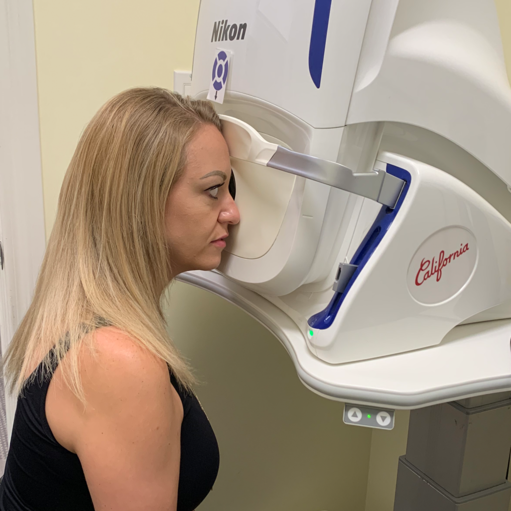
We use cutting-edge digital imaging technology to assess your eyes. Many eye diseases, if detected at an early stage, can be treated successfully without total loss of vision. Your retinal Images will be stored electronically. This gives the eye doctor a permanent record of the condition and state of your retina.

This is very important in assisting your Optometrist to detect and measure any changes to your retina each time you get your eyes examined, as many eye conditions, such as glaucoma, diabetic retinopathy and macular degeneration are diagnosed by detecting changes over time.

The advantages of digital imaging include:

  • Quick, safe, non-invasive and painless
  • Provides detailed images of your retina and sub-surface of your eyes
  • Provides instant, direct imaging of the form and structure of eye tissue
  • Image resolution is extremely high quality
  • Uses eye-safe near-infra-red light
  • No patient prep required

Digital Retinal Imaging allows your eye doctor to evaluate the health of the back of your eye, the retina. It is critical to confirm the health of the retina, optic nerve and other retinal structures. The digital camera snaps a high-resolution digital picture of your retina. This picture clearly shows the health of your eyes and is used as a baseline to track any changes in your eyes in future eye examinations.

Visual Field Testing

A visual field test measures how much ‘side’ vision you have. It is a straightforward test, painless, and does not involve eye drops. Essentially lights are flashed on, and you have to press a button whenever you see the light. Your head is kept still and you have to place your chin on a chin rest. The lights are bright or dim at different stages of the test. Some of the flashes are purely to check you are concentrating.

Each eye is tested separately and the entire test takes 15-45 minutes. Your optometrist may ask only for a driving licence visual field test, which takes 5-10 minutes. If you have just asked for a driving test or the clinic doctor advised you have one, you will be informed of the result by the clinic doctor, in writing, in a few weeks.

Normally the test is carried out by a computerised machine, called a Humphrey. Occasionally the manual test has to be used, a Goldman. For each test you have to look at a central point then press a buzzer each time you see the light.